The Max Pulse-State of the art cardiovascular screening device…

Overview
Cardiovascular & Autonomic Nervous System (ANS) Screening Device
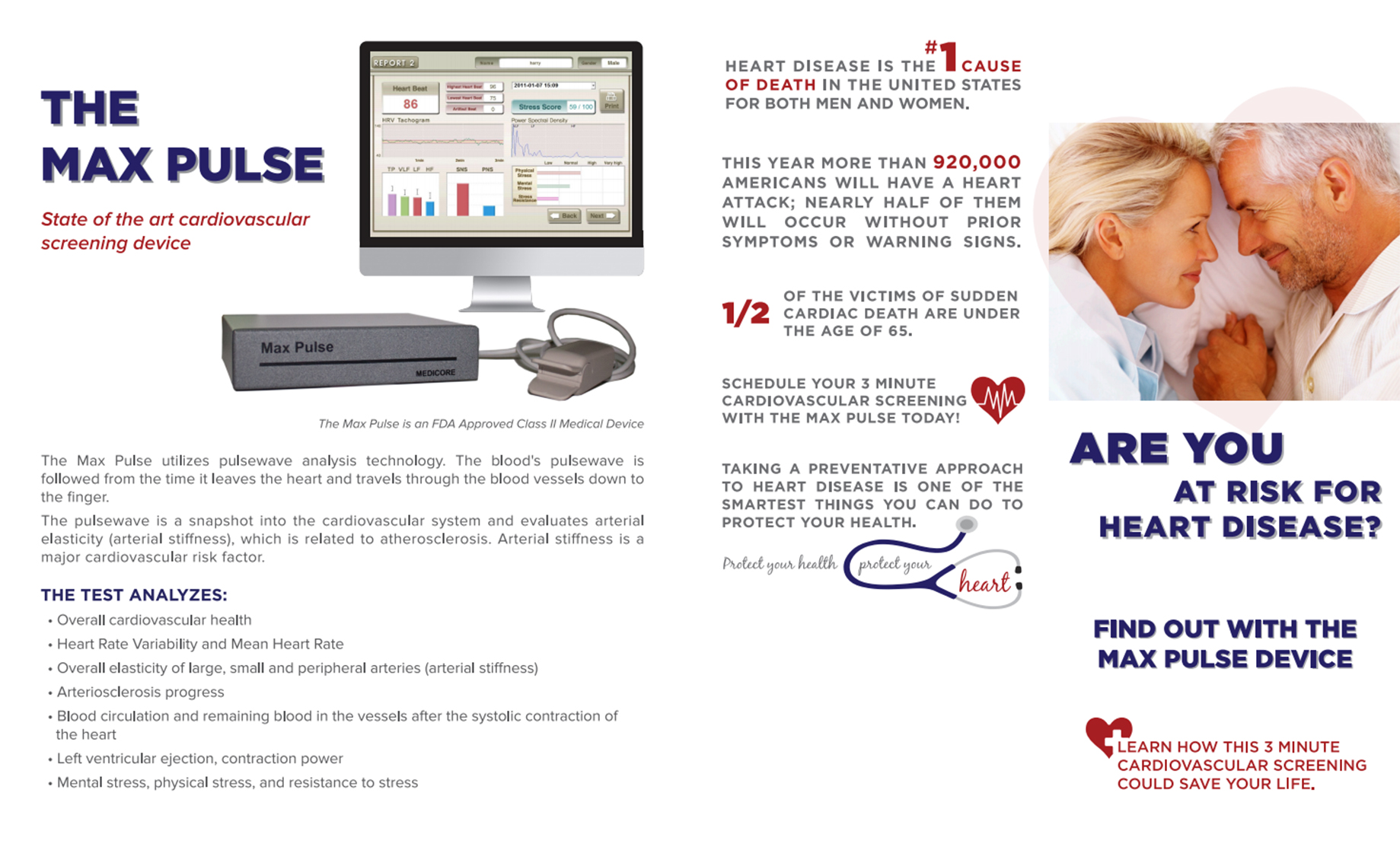
The Max Pulse is a simple, user-friendly, non-invasive, FDA Class II medical screening device. The device provides measurements using Photoelectric Plethysmography, Accelerated Plethysmography, and other technologies to access overall cardiovascular and ANS wellness.
Photoelectric Plethysmography (PTG)
PTG is a non-invasive technique for measuring the amount of blood flow present or passing through, an organ or other part of the body. Plethysmography is used to diagnose deep vein thrombosis and arterial occlusive disease.
Accelerated Plethysmography (APG)
Using a finger clip, the blood’s pulsewave is followed from the time it leaves the heart and travels through the blood vessels down to the finger. The pulsewave is a snapshot into the cardiovascular system and evaluates arterial elasticity (arterial stiffness), which is related to atherosclerosis. Arterial stiffness is a major cardiovascular risk factor.
There is strong scientific evidence supporting the use of plethysmography as a diagnostic and prognostic tool for early warning signs of cardiovascular disease and peripheral vascular disease (including primary and secondary Raynaud’s phenomenon).
THE TEST ANALYZES:
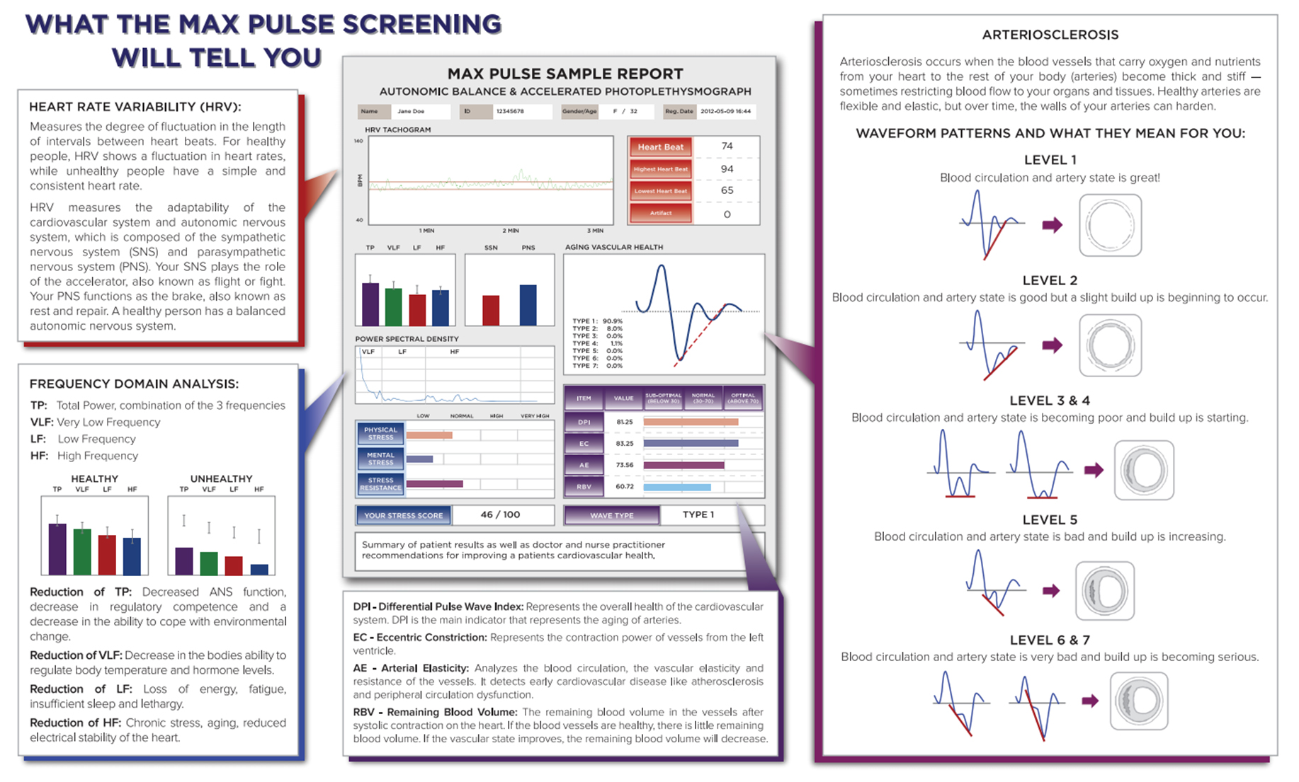
- Heart Rate Variability (HRV) – Determines one’s overall health status and autonomic nerve system. “Meta-analyses of published data demonstrate that reduced cardiovascular autonomic function, as measured by heart rate variability, is strongly associated with an increased risk of silent myocardial ischemia (lack of oxygen to the heart w/o symptoms) and mortality.”
- Differential Pulse Wave Index (DPI) – Overall cardiovascular health.
- Eccentric Constriction (EC) – Constriction power of vessels from the left ventricle.
- Arterial Elasticity (AE) – Overall elasticity of large, small and peripheral arteries (arterial stiffness).
- Remaining Blood Volume (RBV) – Remaining blood in the vessels after systolic contraction of the heart.
- Wave Type – Aging vascular health indicator.
- Mean Heart Rate – Average beats per minute or heart rate.
- Arteriosclerosis Progress – 7 pictorial wave types showing typical artery status.
- Stress Score – Overall stress health compared to resistance levels.
- Stress Levels – Mental stress, physical stress, and resistance to stress. Changes in pressure, velocity, blood volume, and other indices.
- And Other Indices
Max Pulse scanning device is also a useful tool in assisting health-care practitioners, technicians, and individuals in the early detection of cardiovascular related issues. The test will also help assess nutraceutical and/or pharmaceutical needs. Through periodic screenings and lifestyle changes (i.e. exercise, diet, and supplementation etc.), one is able to monitor the effectiveness of these changes and how they relate to the person’s cardiovascular, autonomic and overall health status.
HEART RATE VARIABILITY ANALYSIS
The source information for HRV analysis is continuous beat-by-beat (not averaged) recording of heartbeat intervals. There are many ways to measure and record those intervals. However two such methods are found to be the most appropriate for this.
Pulse wave analysis is way of measuring heartbeat intervals. It is a simple and least invasive method of measurement based on photoplethysmograph (PPG). PPG is a signal reflecting changes in a blood flow in tiny blood vessels typically spotted in fingertips or earlobes. Typical PPG sensor emits infrared light towards the skin area of an earlobe or finger. The blood passing through this area through numerous tiny vessels absorbs certain portion of that light while remaining light is detected by a special photocell. The amount of absorbed light is proportional to the amount of blood passing by. Since the blood flow is not constant due to pulsations caused by heartbeats the sensor generates a very specific waveform reflecting those changes in blood flow. This waveform is usually called as a pulse wave. This waveform can be processed by a special algorithm to derive beat-by-beat heartbeat intervals.
THE CLINICAL MEANING OF A DECREASE IN HRV
It is found that a lowered HRV is associated with aging, decreased autonomic activity, hormonal balance, specific types of autonomic neuropathies (e.g. diabetic neuropathy) and increased risk of sudden cardiac death, after an acute heart attack.
Other research indicates that depression, panic disorders and anxiety have negative impact on autonomic function, typically causing depletion of the parasympathetic tone. On the other hand an increased sympathetic tone is associated with lowered threshold of ventricular fibrillation. These two factors could explain why such autonomic imbalance caused by significant mental and emotional stress increases risk of heart attack followed by sudden cardiac death.
Aside from that, there are multiple studies indicating that HRV is quite useful as a way to quantitatively measure physiological changes caused by various interventions both pharmacological and non-pharmacological during treatment of those conditions manifesting significant reduction in HRV. (See chart 5.3 Diseases Associated with Lowered HRV).
However, it is important to realize, that up to this point in time, the clinical implication of HRV analysis has been clearly recognized in only two medical conditions:
Predictor of risk of arrhythmic events or sudden cardiac death after acute heart attack
Clinical marker of diabetic neuropathy evolution
Nevertheless, as the number of clinical studies involving HRV in various clinical aspects and conditions grows, HRV remains one of the most promising methods of investigating general health in the future.
DISEASES ASSOCIATED WITH LOWERED HRV
- Myocardiac infarction (MI)
- Angina pectoralis
- Ventricular arrhythmia and Premature ventricular contraction (PVC)
- Sudden cardiac death
- Coronary artery disease
- Congestive heart failure
- Diabetes mellitus & Diabetic autonomic neuropathy
- Brain injury
- Epilepsy
- Multiple sclerosis
- Fibromyalgia & Chronic fatigue syndrome
- Obesity
- Guillian-Barre Syndrome
- Depression & Anxiety disorder (Panic disorder)
- Stress induced diseases
DISCLAIMER
For the past 20+ years, methods of the heart rate variability (HRV) analysis have become one of the most popular means of assessment of the autonomic nervous system (ANS) function because of their simple and very informative nature.
At this time there are well-defined standards and methodologies of using methods of HRV analysis, created special normative databases and criteria of assessment of various HRV parameters with regard to their comparison with normative ranges.
At the same time it is very important to point out that there is a tendency in specific cases to over exaggerate diagnostic value of the assessment of results of HRV analysis when professionals attempt to use these results to make conclusions about presence or absence of certain diseases. The Max Pulse scanning device must be used in the scope that it was intended.


